Florida Memory is administered by the Florida Department of State, Division of Library and Information Services, Bureau of Archives and Records Management. The digitized records on Florida Memory come from the collections of the State Archives of Florida and the special collections of the State Library of Florida.

State Archives of Florida
- ArchivesFlorida.com
- State Archives Online Catalog
- ArchivesFlorida.com
- ArchivesFlorida.com
State Library of Florida
Related Sites
Patent for Obstetrical Apparatus.
Date: May 1, 1951
Series: S 900 - Florida State Board of Health Subject files, 1875-1975.
Midwifery.
(Page 2 of 3)
Early Florida Medicine
Transcript
[page 2]
2,551,433
3
[left column]
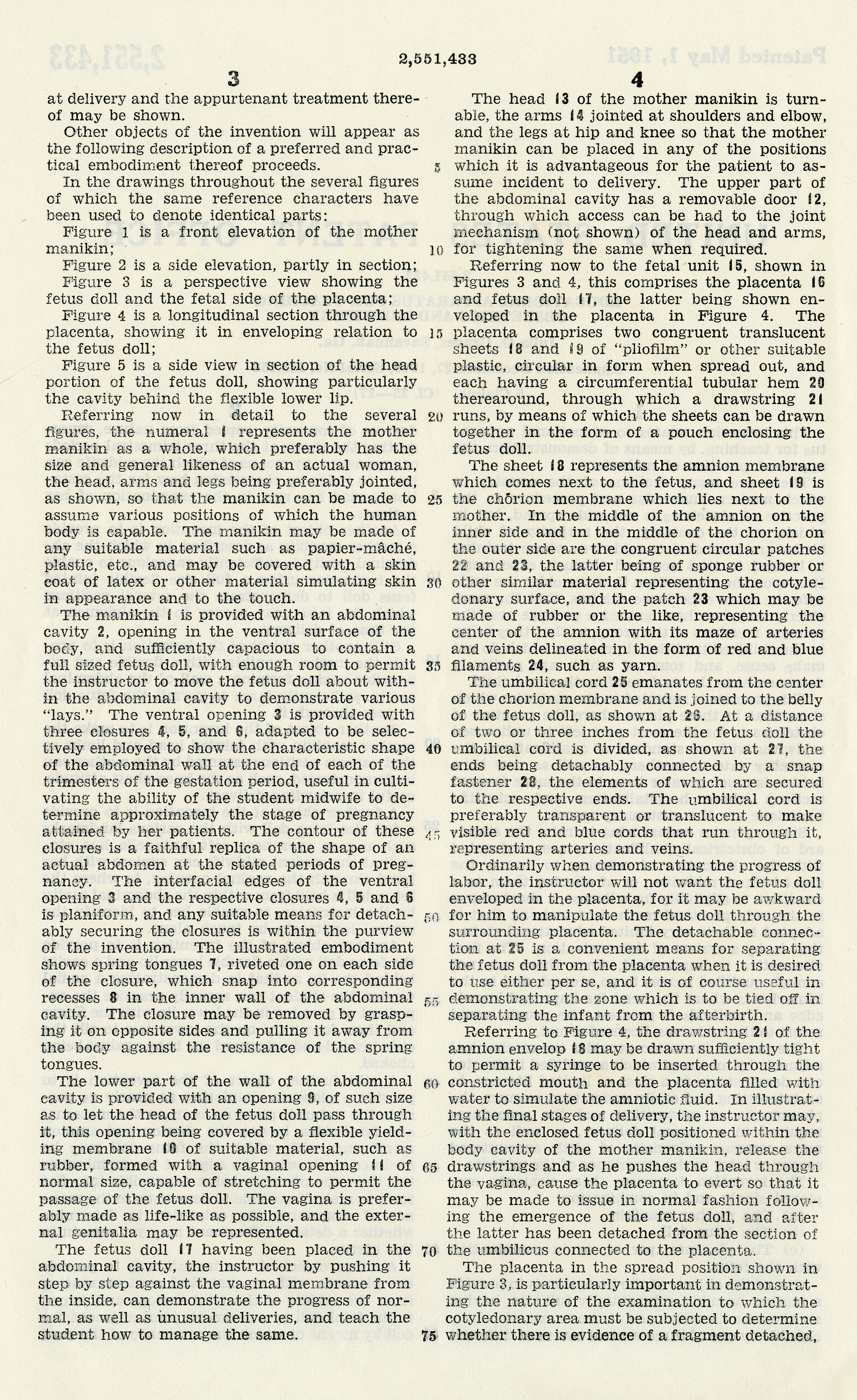
at delivery and the appurtenant treatment there-
of may be shown.
Other objects of the invention will appear as
the following description of a preferred and prac-
tical embodiment thereof proceeds. 5
In the drawing throughout the several figures
of which the same reference characters have
been used to denote identical parts:
Figure 1 is a front elevation of the mother
manikin; 10
Figure 2 is a side elevation, partly in section;
Figure 3 is a perspective view showing the
fetus doll and the fetal side of the placenta;
Figure 4 is a longitudinal section through the
placenta, showing it in enveloping relation to 15
the fetus doll;
Figure 5 is a side view in section of the head
portion of the fetus doll, showing particularly
the cavity behind the flexible lower lip.
Referring now in detail to the several 20
figures, the numeral 1 represents the mother
manikin as a whole, which preferably has the
size and general likeness of an actual woman,
the head, arms and legs being preferably jointed,
as shown, so that the manikin can be made to 25
assume various positions of which the human
body is capable. The manikin may be made of
any suitable material such as papier-mâché,
plastic, etc., and may be covered with a skin
coat of latex or other material simulating skin 30
in appearance and to the touch.
The manikin 1 is provided with an abdominal
cavity 2, opening in the ventral surface of the
body, and sufficiently capacious to contain a
full sized fetus doll, with enough room to permit 35
the instructor to move the fetus doll about with-
in the abdominal cavity to demonstrate various
"lays." The ventral opening 3 is provided with
three closures 4, 5, and 6, adapted to be selec-
tively employed to show the characteristic shape 40
of the abdominal wall at the end of each of the
trimesters of the gestation period, useful in culti-
vating the ability of the student midwife to de-
termine approximately the stage of pregnancy
attained by her patients. The contour of these 45
closures is a faithful replica of the shape of an
actual abdomen at the stated periods of preg-
nancy. The interfacial edges of the ventral
opening 3 and the respective closures 4, 5, and 6,
is planiform, and any suitable means for detach- 50
ably securing the closures is within the purview
of the invention. The illustrated embodiment
shows spring tongues 7, riveted one on each side
of the closure, which snap into corresponding
recesses 8 in the inner wall of the abdominal 55
cavity. The closure may be removed by grasp-
ing it on opposite sides and pulling it away from
the body against the resistance of the spring
tongues.
The lower part of the wall of the abdominal 60
cavity is provided with an opening 9, of such size
as to let the head of the fetus doll pass through
it, this opening being covered by a flexible yield-
ing membrane 10 of suitable material, such as
rubber, formed with a vaginal opening 11 of 65
normal size, capable of stretching to permit the
passage of the fetus doll. The vagina is prefer-
ably made as life-like as possible, and the exter-
nal genitalia may be represented.
The fetus doll 17 having been placed in the 70
abdominal cavity, the instructor by pushing it
step by step against the vaginal membrane from
the inside, can demonstrate the progress of nor-
mal, as well as unusual deliveries, and teach the
student how to manage the same. 75
[right column]
4
The head 13 of the mother manikin is turn-
able, the arms 14 jointed at shoulders and elbow,
and the legs at hip and knee so that the mother
manikin can be placed in any of the positions
which it is advantageous for the patient to as-
sume incident to delivery. The upper part of
the abdominal cavity has a removable door 12,
through which access can be had to the joint
mechanism (not shown) of the head and arms,
for the tightening the same when required.
Referring now to the fetal unit 15, shown in
Figures 3 and 4, this comprises the placenta 16
and fetus doll 17, the latter being shown en-
veloped in the placenta in Figure 4. The
placenta comprises two congruent translucent
sheets 18 and 9 of "pliofilm" or other suitable
plastic, circular in form when spread out, and
each having a circumferential tubular hem 20
therearound, through which a drawstring 21
runs, by means of which the sheets can be drawn
together in the form of a pouch enclosing the
fetus doll.
The sheet 18 represents the amnion membrane
which comes next to the fetus, and sheet 19 is
the chorion membrane which lies next to the
mother, In the middle of the amnion on the
inner side and in the middle of the chorion on
the outer side are the congruent circular patches
22 and 23, the latter being of sponge rubber or
other similar material representing the cotyle-
donary surface, and the patch 23 which may be
made of rubber or the like, representing the
center of the amnion with its maze of arteries
and veins delineated in the form of red and blue
filaments 24, such as yarn.
The umbilical cord 25 emanates from the center
of the chorion membrane and is joined to the belly
of the fetus doll, as shown at 26. At a distance
of two or three inches from the fetus doll the
umbilical cord divided, as shown at 27, the
ends being detachably connected by a snap
fastener 28, the elements of which are secured
to the respective ends. The umbilical cord is
preferably transparent or translucent to make
visible red and blue cords that run through it,
representing arteries and veins.
Ordinarily when demonstrating the progress of
labor, the instructor will not want the fetus doll
enveloped in the placenta, for it may be awkward
for him to manipulate the fetus doll through the
surrounding placenta. The detachable connec-
tion at 25 is a convenient means for separating
the fetus doll from the placenta when it is desired
to use either per se, and it is of course useful in
demonstrating the zone which is to be tied off in
separating the infant from the afterbirth.
Referring to Figure 4, the drawstring 21 of the
amnion envelop 18 may be drawn sufficiently tight
to permit a syringe to be inserted through the
constricted mouth and the placenta filled with
water to simulate the amniotic fluid. In illustrat-
ing the final stages of delivery, the instructor may,
with the enclosed fetus doll positioned within the
body cavity of the mother manikin, release the
drawstrings and as he pushes the head through
the vagina, cause the placenta to evert so that it
may be made to issue in normal fashion follow-
ing the emergence of the fetus doll, and after
the latter has been detached from the section of
the umbilicus connected to the placenta.
The placenta in the spread position shown in
Figure 3, is particularly important in demonstrat-
ing the nature of the examination to which the
cotyledonary area must be subjected to determine
whether there is evidence of a fragment detached,
Features
Territorial Legislative Council Records, 1822-1845
Documenting Florida's legislature prior to statehood
State and County Officer Directories, 1868-1969
Lists of Florida's state and county officials either elected or appointed between 1868 and 1969.
Guide to Military Records and the Wartime Experience
A guide to government records and manuscript collections relating to service members and the wartime experience.
Dust Tracks
21 tracks from Hurston's time documenting African American culture for the WPA in 1930s Florida.
History Day 2025
Resources for Florida History Day from the State Library and Archives of Florida.